Does Ultrasonography Help Detect Hormonal Changes?
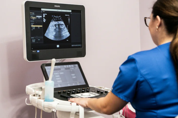
Photo: adoria.lv
Ultrasonography has become one of most widely used thus indispensable visual diagnostic methods in modern medicine providing opportunity safely without invasive intervention in real time give detailed insight into human body's internal organ structure. Hormonal changes in turn complex processes significantly affecting various body functions overall health condition.
How ultrasonographic examination can help identify organ structural changes potentially indicating possible hormonal disorders how informative in this context ultrasound for pregnant women – continues to tell Health and Beauty Center gynecologist Jana Bjornsone.
Ultrasonography Hormonal Changes – Summary:
- USG hormones: ultrasonography itself doesn't determine hormone levels but can help detect organ structural changes potentially early indicating possible hormonal disorders.
- Diagnostic value: method helps detect structural changes in ovaries uterus other organs potentially signaling hormonal imbalance.
- Pregnancy context: ultrasound for pregnant women essential both in pregnancy planning course assess hormonal process impact.
Ultrasonography's Role in Hormonal Process Assessment
Ultrasonographic examinations themselves don't directly determine hormone levels in blood – for this purpose specific laboratory tests intended. However ultrasonography invaluable visual diagnostic method as with its help doctor can detailed view assess various organs' shape size internal structure.
As hormone balance affects many of our organs' health ultrasonography-noticed changes can serve as early warning signals indicating possible problems in hormone system function.
- More about what gynecological ultrasonography what cases needed what information provides about woman's reproductive health read article: "What Gynecological Ultrasonography When Needed?".
Women's Reproductive System Assessment
Woman's reproductive system one of most illustrative examples where ultrasonography provides particularly much information about hormonal process course as both ovaries uterus condition very closely dynamically related to hormone fluctuations not only during monthly cycle but also in other significant life stages.
Performing ovarian ultrasonography specialist can carefully evaluate their size internal structure also follicle – small blisters where eggs mature – number development stage. Exactly these observations help understand ovarian functional state as their activity directly regulated by such important hormones as estrogen progesterone follicle-stimulating hormone (FSH). These hormones' coordinated action crucial both for regular menstrual cycle woman's ability conceive.
No less important uterine ultrasonographic examination where special attention paid to its lining endometrium. Endometrium also cyclically changes under hormone influence each month becoming thicker preparing for possible pregnancy. Endometrium condition essential both for normal menstruation course embryo successful implantation in uterus further development after fertilization. Thus ultrasonography helps assess whether these natural hormone-guided processes in woman's body proceed correctly.
Ultrasonography During Menopause Period
Menopause natural process in woman's life characterized by significant hormonal changes mainly ovarian function exhaustion estrogen level reduction. Also in this period ultrasonography has important role as allows assess changes in uterus ovaries related to this new hormonal background.
E.g., ovaries during menopause usually decrease in size no longer follicle maturation occurs meanwhile uterine lining (endometrium) should be thin. Ultrasonography helps timely notice any atypical changes e.g., endometrium thickening potentially indicating pathological processes requiring additional examination also assess ovarian condition.
Thus also during menopause preventive USG examinations very important monitor woman's health timely respond to possible problems arising as hormonal balance changes.
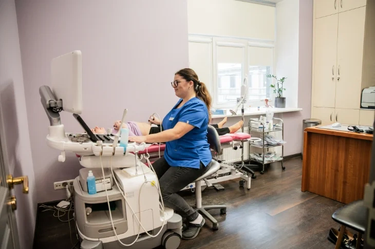
Photo: adoria.lv
Other Organs Diagnostic Indications
Ultrasonography's importance in hormonal process evaluation doesn't limit only to reproductive system. E.g., with its help can effectively examine also thyroid – small but very important organ producing hormones regulating almost all processes in body.
In ultrasonography can timely notice thyroid structure changes nodules potentially indicating function disorders thus – for broader hormonal balance problems.
Overall sometimes image seen in ultrasonography can be so characteristic of some specific health condition helps direct doctor's diagnostic thinking plan further steps. In such cases ultrasonography results serve as important signal prompting perform additional checks e.g., hormone blood tests clarify diagnosis select most appropriate further action.
Hormonal Changes Ultrasonography for Pregnant Women: From Planning to Postpartum Period
Ultrasonography important examination accompanying woman in all pregnancy stages – from conception planning to postpartum period helping understand hormonal change impact. Let's look at what main ultrasonography tasks in each these stages.
Ultrasonography in Pregnancy Planning Stage
In pregnancy planning stage ultrasonography has crucial role assess woman's reproductive system condition identify possible obstacles to conception potentially related to hormonal factors. Main ultrasonography tasks in this period include:
- ovulation disorder diagnostics: performing folliculometry ultrasonographic ovarian monitoring during menstrual cycle possible follow follicle growth precisely determine ovulation course. Particularly important if infertility suspected;
- hormonal disorder identification: such conditions as polycystic ovary syndrome (PCOS) characterized by specific ovarian structure changes e.g., many small follicles enlarged ovaries can be diagnosed confirmed. This information very important for further treatment tactics selection;
- other reproductive system feature assessment: e.g., possible diagnose uterine anomalies fibroids endometrium pathologies also affecting successful pregnancy onset.
Ultrasonography's Importance During Pregnancy
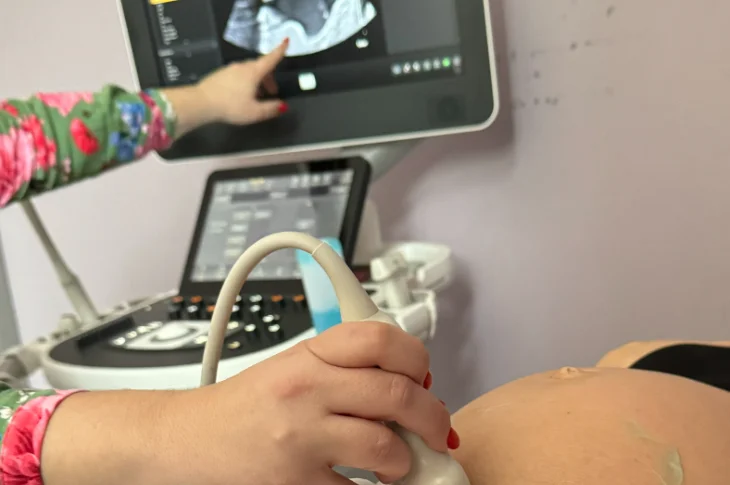
Photo: adoria.lv
When pregnancy occurs woman's body undergoes significant hormonal changes vital for normal fetal development pregnancy course. Ultrasound for pregnant women one of main safest ways follow these processes. Though USG doesn't directly measure hormone levels provides valuable indirect information about hormonal system function allowing assess:
- placenta condition: its structure maturity degree attachment site dependent on hormonal balance essential for fetal supply with nutrients;
- corpus luteum cyst: in early pregnancy ovary forms corpus luteum appearing as cyst in USG producing progesterone – hormone maintaining pregnancy in its early stage.
What checks examinations recommended for pregnant woman in first pregnancy weeks? Learn reading our specialists prepared article: "First Weeks in Pregnancy: What Checks Perform?".
Full pregnant women care includes several routine examinations quality ultrasound for pregnant women indispensable part of these checks in different pregnancy stages. These examinations' main goals:
- fetal growth development assessment: examinations ensure baby growing according to pregnancy time;
- congenital anomalies timely detection: early diagnostics allows plan necessary care;
- fetal well-being assessment: movements heart rate amniotic fluid amount evaluated;
- placenta localization function check: important rule out problems like placenta previa;
- indirect indications of mother's health problems: e.g., if prospective mother develops gestational diabetes ultrasonography can observe larger fetus (macrosomia) potentially indicating this hormonal imbalance.
Ultrasonography in Postpartum Period
Also after baby's birth ultrasonography can have important role in woman's health assessment as body experiences new hormonal changes returning to pre-pregnancy state. In postpartum period ultrasonography usually performed if specific complaints suspicion of complications. Helps:
- assess uterine involution (contraction): check whether uterus contracts quickly enough appropriately hormone-guided process. Slow contraction can indicate problems;
- rule out retained placenta parts: ensure no placenta fetal membrane parts remained in uterus potentially causing bleeding infection;
- diagnose postpartum complications: detect possible hematomas (hemorrhages) infection signs in uterus other complications;
- check other pelvic organs condition if necessary: also assess ovaries bladder condition after birth.
Such examination helps ensure woman's recovery after birth proceeds successfully without complications.
Modern Health Care at Health and Beauty Center Adoria

Photo: adoria.lv
Gynecology in Riga Health and Beauty Center Adoria A. Čaka street 70-3 – modern individual reproductive health care for women in all life stages state-funded pregnant women care precise USG examinations with latest generation equipment. Also provide wide range of general health care services for entire family including wide spectrum of USG examinations. Book visit now!