Is a 3D dental X-ray harmful to pregnant women and children?
Photo: freepik.com/Freepik
Quality dental care today is unthinkable without precise examinations, yet the question of whether X-rays are harmful still worries many people. This is especially relevant for expectant mothers and parents who fear the effect of ionising radiation on a child's body or on the development of the foetus.
In reality, timely diagnostics are an essential prerequisite for safe treatment, which is why the dental 3D X-ray has become a standardised diagnostic method in modern dentistry. It makes it possible to detect hidden inflammatory processes in time and to prevent complications whose treatment, without precise visual data, would be imprecise or even impossible.
Adoria Health and Beauty Centre dentist Ieva Breča explains in the rest of this article what the real radiation doses are and why there is no reason to fear 3D digital examinations
In this article you will learn:
- Safety standards: why the modern dental 3D X-ray is safe even for the most sensitive groups of patients.
- Pregnancy specifics: what precautions are taken when performing a dental X-ray on pregnant women.
- Children's diagnostics: why a timely X-ray for a child is less dangerous than an untreated dental infection.
Why is digital X-ray diagnostics the safest choice today?
Many patients' concerns about radiation are rooted in their experience with outdated, analogue equipment, but in modern medicine the situation has fundamentally changed. The traditional 2D image is still used for general examination, but the low-radiation digital X-ray has significantly reduced the required exposure dose. For comparison – the radiation from a single digital tooth image is so negligible that it is equivalent to the natural background radiation each of us receives from the surrounding environment in just a couple of hours.
The 3D X-ray, in turn, raises diagnostics to a new level, providing a spatial visualisation of the jaw with minimal impact on the body. The safety and precision of this technology are guaranteed by several essential factors.
- Focused beam: unlike general medical examinations, dental equipment focuses the beam precisely on the area being examined, without emitting unnecessary energy onto the rest of the body.
- High-sensitivity sensors: modern digital sensors are incomparably more sensitive than the films used in the past, so obtaining a quality image requires a considerably smaller amount of radiation.
- Individual protection: even at minimal doses, lead aprons and collars are used, which fully protect the most sensitive organs, such as the thyroid gland, from any effect of scattered radiation.
- Low exposure time: digital data processing happens in an instant, which means the patient is exposed to the rays for only a few seconds.
How to understand the units of radiation dose and their significance?
To objectively answer the question of whether X-rays are harmful, we must look at the units in which the effect of radiation on tissue is measured – microsieverts (µSv). The average natural background radiation that everyone receives from the sun and the earth is approximately 3000 µSv per year. A single digital dental X-ray delivers only about 1–5 µSv, which is a negligible fraction of the annual natural background. Even a more complex dental 3D X-ray, which provides extensive information about all the jaw tissues, does not exceed safety norms and is comparable to a flight across the ocean.
This precise information helps the doctor see even the smallest changes in bone tissue, the location of nerves and the condition of the paranasal sinuses. Digital technologies make it possible to process the image on a computer, enlarge it and change the contrast, so it is no longer necessary to take repeat images if the first one is not clear enough.
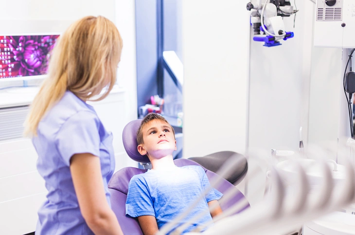
Photo: freepik.com/Freepik
Does a dental X-ray really endanger the foetus in pregnant women?
The medical literature is dominated by the view that a dental X-ray poses no direct threat to the foetus in pregnant women, provided strict protection protocols are followed. Unlike older-generation equipment, modern digital examinations focus the radiation only on the jaw area, and a lead apron fully isolates the rest of the body.
However, the effect of radiation is always assessed in proportion to the benefit – while a regular 2D X-ray provides a flat image with possible overlapping distortions, the modern dental 3D X-ray provides millimetre precision. This is decisive in avoiding erroneous manipulations or receiving a repeat radiation dose due to imprecise data.
It is essential to be aware that an untreated tooth root inflammation or pocket of pus poses an incomparably greater risk than the diagnostics itself. During pregnancy, the oral cavity becomes especially vulnerable not only due to hormonal fluctuations but also due to physiological changes, so regular visits and consultations with a gynaecologist must be supplemented with preventive visits to the dentist and dental hygienist.
Under the influence of hormones, the pH level and viscosity of saliva change, reducing the natural remineralisation capacity of the enamel and promoting the development of caries. Likewise, frequent nausea and vomiting in the first trimester create an acidic environment that chemically erodes the tooth enamel (erosion), making teeth fragile and sensitive.
In cases where an acute oral infection is found, a dental 3D X-ray examination makes it possible to precisely localise the focus of inflammation and begin targeted treatment. This is essential, because untreated, chronic inflammations in the oral cavity can become a permanent source of infection for the whole body, which in some studies is associated with elevated levels of systemic inflammatory markers, thus potentially also affecting the course of pregnancy.
Photo: adoria.lv
Why is an X-ray important for timely diagnostics in a child?
Parents often worry about the effect of radiation, yet modern children's dentistry is unthinkable without precise visualisation methods that make it possible to notice such hidden damage as caries between the teeth or bite deviations at the very beginning, when the child still has no complaints.
It is precisely modern 3D X-ray technology that dispels these concerns, providing an extremely fast and safe examination in which obtaining the image takes only a few seconds, thus causing the child no unnecessary stress. Without this method, the specialist cannot fully assess the condition of the tooth buds, which is decisive in correcting bite formation in time and avoiding lengthy orthodontic treatment in the future.
This approach makes it possible to begin gentle therapy at an early stage, protecting the child from unpleasant sensations and creating a positive, trust-filled experience with the specialist. Since an X-ray performed in time gives a comprehensive picture of the child's oral health, parents can be confident in a precise treatment plan. Modern dental diagnostics in childhood provides several essential advantages:
- Checking tooth buds: the ability to make sure that the permanent teeth are forming correctly and that there is enough room in the jaw for them to grow.
- Examining the spaces between teeth: diagnosing caries in places where teeth touch and where damage is not visible in a regular examination.
- Monitoring jaw development: timely detection of an incorrect bite, making it possible to begin correction at the most suitable time for the child.
- Preventing hidden infections: the ability to find inflammations in time that cause no pain but can seriously damage the enamel of the developing permanent teeth.
Using modern dental 3D X-ray technologies, the radiation dose is individually adapted to the child's height and weight, ensuring maximum safety and a medically justified result.
Frequently asked questions about dental X-rays for pregnant women and children
Below we have compiled answers to the questions that patients most often ask before undergoing digital diagnostics, to help better understand the safety and necessity of the procedure.
- How often can a dental X-ray be taken without risk to health?
For preventive purposes, it is recommended that adults have an X-ray once every 1–2 years, while for children the interval is adapted to caries risk and the phases of jaw development. During treatment, for example in the case of endodontics or implantology, the examination can be performed more often, because with modern digital technology the cumulative radiation dose, even with several images per year, remains far below the permissible medical norms. The main indicator is medical usefulness – each examination is performed only when it provides essential information for precise treatment.
- Can breastfeeding mothers undergo X-ray examinations?
Yes, breastfeeding mothers have no restrictions on having a dental X-ray. The ionising radiation used in diagnostics does not accumulate in breast milk and in no way affects its biological composition or quality. Unlike some medical preparations, X-rays pass through tissue in an instant and leave no trace in the body, so breastfeeding can be safely continued immediately after the visit to the specialist.
- Is a dental 3D X-ray mandatory before every caries treatment?
For ordinary, superficial caries treatment or filling, a three-dimensional examination is not mandatory, but it becomes indispensable if there is a suspicion of hidden processes. A dental 3D X-ray is the mandatory standard before complex operations, root canal treatment, or when the extent of inflammation cannot be precisely determined by visual examination and a 2D image. It allows the specialist to see the tooth from all sides, protecting the patient from unforeseen complications and ensuring maximum longevity of the filling or restoration.
Take care of your dental health safely at Adoria Health and Beauty Centre!
Photo: adoria.lv
Maintaining dental health is important at any stage of life, and modern technology makes it possible to do so without unnecessary risk to health. Adoria Health and Beauty Centre provides the highest-quality dental 3D X-ray examinations and a wide range of dental services for the whole family. Our specialists will take care of your well-being, offering safe and professional care for both you and your children. Book a visit by calling +371 67 315 000 or by filling out the application form on our website, and see for yourself that modern dentistry is a comfortable and safe choice.