Hidden dental problems: what does the doctor see on a panoramic X-ray?
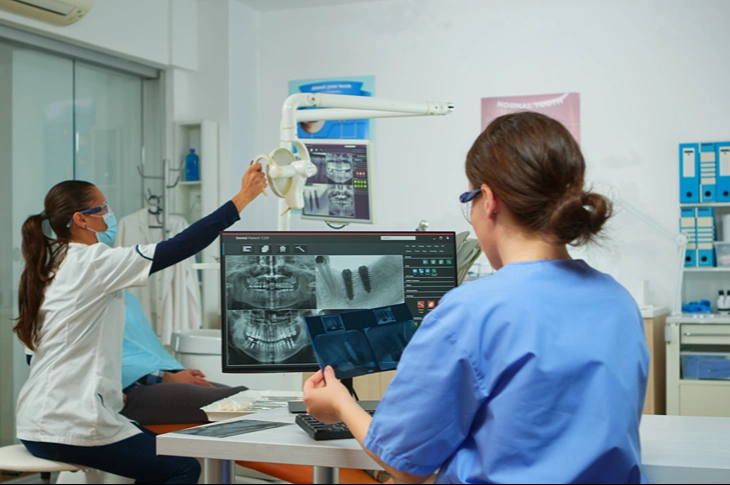
Photo: freepik.com/Freepik
Modern dentistry is unthinkable without precise diagnostics, which make it possible to see processes that remain hidden from the human eye during a visual examination. The most advanced diagnostic tool currently available for a comprehensive assessment of oral cavity health is the panoramic X-ray, which provides a detailed and broad overview of the jaw system as a whole.
Polina Bite, a dentist at the Adoria Health and Beauty Centre, explains in more detail in the following article what purposes this method is used for and which pathologies it helps to detect in good time
In this article you will learn:
- The benefits of diagnostics: what hidden problems are revealed by a full overview image of the jaws.
- Detecting inflammations: why this examination is the best way to diagnose chronic processes.
- Planning a visit: in which cases the specialist decides that this imaging method is necessary.
Why is a panoramic X-ray the basis for accurate dental health diagnostics?
Although small dental images are useful for detecting caries in individual teeth, a panoramic X-ray offers a complete view of the entire oral cavity in a single image. This examination, which in medicine is also called an orthopantomogram, covers both jaws, all the teeth, the paranasal sinuses and the jaw joints. Imaging on this scale is irreplaceable, because many serious health problems begin asymptomatically and cause no pain until the process has already become chronic or has caused tissue damage.
Digital technology provides high image resolution while using a minimal radiation dose. By analysing this image, the dentist can assess not only the condition of the existing teeth, but also the density of the bone tissue and the anatomical features of the surrounding structures. This is especially important in cases where complex treatment planning or a pre-surgical assessment is required. Diagnostics carried out in good time helps to avoid unforeseen complications during procedures and allows the specialist to prepare for specific anatomical nuances that would otherwise remain unknown.
Photo: adoria.lv
What does the doctor see on a panoramic X-ray image?
A panoramic image allows the specialist to assess the health of the oral cavity as a whole, noticing things that are not visible during an ordinary examination. It provides a clear picture of the processes taking place beneath the gums and in the jawbone, making it possible to prevent complications in time.
- Inflammations and formations: makes it possible to notice chronic processes at the tooth roots, as well as various cysts or other unhealthy processes in the bone.
- The condition of tooth roots: provides information about the shape and number of the roots and how close they are to the jaw nerves or the paranasal sinuses.
- Hidden damage: helps to spot caries in places where it is not visible to the eye, for example under existing fillings or crowns.
- Bone tissue health: makes it possible to assess whether periodontitis has begun and to what extent it has affected the supporting tissues of the tooth along the entire length of the jaw.
- The position of wisdom teeth: shows whether they are growing correctly and not endangering the neighbouring teeth.
- Peculiarities in the number of teeth: reveals extra tooth buds or situations where a tooth has not formed at all.
Since many processes in the tissues take place unnoticed, regular professional dental hygiene is the best way to keep oral cavity health under control. In that case, a panoramic image prescribed by the specialist is no longer a necessity for rescuing a neglected situation, but rather a reliable monitoring mechanism that confirms that all the teeth and the surrounding bone tissue are healthy.
Why is it important to detect a dental cyst in time?
Many patients have no idea that a dental cyst is developing in their jawbone, because it may cause no unpleasant sensations for years. A cyst is a pathological formation, usually filled with fluid, that slowly expands, pushing aside and destroying healthy bone. Often it arises at the tip of a tooth root as a consequence of an untreated canal inflammation or an injury.
If a cyst is not detected in time on a panoramic X-ray, it can reach a considerable size, causing the jawbone to weaken or even leading to spontaneous fractures. Early diagnosis of cysts makes it possible to carry out more conservative surgical manipulations, preserving as much healthy tissue as possible. Once the formation has been precisely diagnosed, the doctor plans the treatment using gentle methods that protect the surrounding healthy tissue as much as possible.
Such an approach ensures a faster recovery period and prevents the risk that the inflammation could spread to the neighbouring teeth or to the paranasal sinuses. 3D digital dental diagnostics is the first step towards preventing the serious consequences caused by such processes, which are initially often asymptomatic but destroy tissue.
What problems can a chronic inflammation under a tooth conceal?
Photo: adoria.lv
A chronic inflammation under a tooth, or a granuloma, is one of those pathologies for the identification of which a panoramic X-ray is especially useful. This situation indicates that the infection has spread beyond the tooth's root canals and has begun to affect the surrounding bone tissue. Although such a focus may cause no complaints for years, it serves as a constant source of infection in the body, which not only burdens the immune system but can at any moment turn into an acute stage, causing severe pain and swelling. A precise digital image allows the specialist to make well-founded decisions about further treatment, providing the opportunity to:
- Determine the extent of the inflammation: assess the size of the focus and its boundaries in the jawbone.
- Analyse previous treatment: check the quality of the root canal filling if the tooth has already been treated.
- Assess the possibilities of preserving the tooth: make an objective decision – whether the tooth can be saved, or whether the better choice is to extract it.
- Minimise risks to neighbouring structures: see precisely how close the infection is to the jaw nerve or the paranasal sinuses.
If, during diagnostics, the dental surgeon discovers that the inflammation has affected too large an area and that therapeutic canal cleaning no longer guarantees that the tooth can be saved, the specialist may recommend surgical removal of the inflammation focus or, in some cases, extraction of the tooth. In such cases, dental surgery makes it possible to eliminate the source of infection gently but completely, preventing further destruction of the bone tissue and protecting the patient's general health from the risks of systemic inflammation.
In which cases is it important to take a panoramic X-ray for wisdom teeth?
When planning the removal of the eighth teeth, or wisdom teeth, 3D digital X-ray diagnostics gives the doctor critically important information about their actual position in the jaw. These teeth are often located in complicated positions – they may be crooked, fully or partially trapped in the bone (impacted), or even growing horizontally against the neighbouring teeth. That is why a panoramic image is an irreplaceable tool for allowing the specialist to clearly see the anatomy of the roots and their precise location in relation to the jaw canal in which the main nerve is situated.
A digital X-ray for wisdom teeth allows the specialist to prepare for the procedure by anticipating the curves of the roots and their contact with the neighbouring anatomical structures even before manipulations begin. Such visualisation helps to minimise possible risks and provides several essential advantages:
- Contact with nerve canals: the possibility of assessing how close the tooth roots are to the lower jaw nerve, in order to plan a safe extraction and avoid sensory disturbances in the lip or chin area.
- Impact on neighbouring teeth: a check of whether the wisdom tooth is putting pressure on the seventh tooth, which can promote enamel damage or cause damage (resorption) to the root of the neighbouring tooth.
- The position of stuck teeth: precise diagnostics for impacted teeth that are unable to erupt on their own and often become a source of chronic inflammation in the bone.
- Orthodontic prognosis: for children and adolescents, this image helps to predict whether there will be enough room for the wisdom teeth, so that their growth does not cause the front teeth to shift or lead to bite problems.
How does a panoramic X-ray help to prepare for further treatment?
Photo: adoria.lv
Before starting any extensive treatment, the specialist needs to see the overall picture in order to create a logical and medically justified treatment plan. A panoramic X-ray serves as a "map" of the patient's oral cavity health, on which all the critical zones are marked. This is especially important before prosthetics or implantation, in order to make sure that there are no hidden foci of infection in the bone that could endanger the stability and longevity of the new construction.
Modern 3D dental X-ray diagnostics provides several advantages in the treatment process:
- Precise planning: the possibility of setting priorities – which problems are acute and which require long-term monitoring.
- A unified view: information about the soft and hard tissues, which helps to better understand the patient's individual anatomy.
- Monitoring results: the possibility of comparing images before and after treatment in order to follow the bone tissue regeneration process.
- Effective communication: by seeing the large-format image on the computer screen, the patient better understands the specialist's explanations about the necessary manipulations.
Frequently asked questions about the panoramic X-ray examination
Below we have gathered answers to the questions that patients most often ask before having a panoramic X-ray, in order to help better understand the significance and safety of this method.
- How often is it necessary to take a panoramic X-ray for preventive purposes?
For the majority of adults, it is recommended to take such an overview image once every two years, in order to detect asymptomatic changes in the jawbone or around the tooth roots in good time. If the patient has chronic gum diseases, such as periodontitis, or extensive reconstructive treatment is planned, the specialist may prescribe the examination more often.
- Can a panoramic X-ray completely replace small dental X-rays?
Although a panoramic X-ray provides an excellent general overview, it does not replace small (intraoral) images when maximum detail is needed for diagnosing caries in one specific tooth. Small images focus on precisely depicting the enamel and the interdental surfaces, whereas the panoramic method is intended for seeing the overall picture – the jaw joints, the sinuses and the general arrangement of the teeth. Often both methods complement each other so that the doctor can create a 100% safe and accurate treatment plan.
- Is this examination suitable for children before orthodontic treatment?
Yes, in childhood this examination is important for assessing the presence of permanent tooth buds and the direction of their eruption. Diagnostics carried out in good time allows the orthodontist to notice a potential lack of space in the jaw or an incorrect bite forming even before the first visible problems appear. Likewise, digital visualisation helps to monitor the development of the wisdom teeth, which in adolescence can begin to affect an already aligned row of teeth.
Why choose the Adoria Health and Beauty Centre for diagnostics?

Photo: adoria.lv
At the Adoria Health and Beauty Centre dental clinic in Riga, at A. Čaka iela 70–3, a modern digital panoramic X-ray is available, guaranteeing high precision in the treatment process. Whether you need preventive dental hygiene, caries treatment or extensive dental surgery, the modern equipment allows us to see the tiniest nuances and to prevent problems even before they begin to cause discomfort. Book an appointment by calling +371 67 315 000 or by filling in the application on the website, and make sure that professional dentistry is the best investment in your health!