Preparing for dental implantation: why is a 3D examination necessary?

Foto: freepik.com/Freepik
Tooth loss is not only an aesthetic defect, but also a serious functional impairment that over time can affect both self-confidence and overall health. Modern dentistry offers effective solutions for restoring the smile, and one of the most popular is the use of artificial roots, or implants. However, to achieve a quality result, careful preparation for dental implantation is a critically important first step. It makes it possible to ensure that the new tooth will be stable and serve fully for decades.
Why it is important to carry out an in-depth three-dimensional examination before placing the implant, and how such diagnostics help to avoid the risk of complications, is described in the continuation of the article by the dentist of the Adoria Health and Beauty Centre Darja Gvergžde.
In this article you will learn:
- The advantages of 3D diagnostics: why a traditional 2D X-ray is not sufficient for the precise positioning of the implant.
- The condition of the bone tissue: how a digital examination helps to determine the bone density and the need for additional procedures.
- Reducing risks: which anatomical structures are protected thanks to prior three-dimensional planning.
Why is a traditional panoramic X-ray not sufficient in implantology?
When planning the placement of dental implants, the specialist needs absolute precision, because every millimetre in the anatomy of the oral cavity is decisive. A traditional panoramic X-ray, or orthopantomogram, provides only a two-dimensional image, which often creates optical distortions and does not allow the thickness of the jawbone to be assessed.
That is precisely why preparation for dental implantation includes cone beam computed tomography (CBCT), which creates a virtual model of the jaw. This technology allows the doctor to virtually "walk through" the patient's jaw, seeing not only the height of the bone, but also its volume in space. Without such data, the placement of dental implants would become an unpredictable process, because only in a 3D image is it possible to:
- Assess the quality of the bone: precisely measure the width of the bone at the site where the titanium screw is planned to be placed.
- Localise the nerves: see the precise location of the lower jaw nerve canal, in order not to traumatise it during the procedure.
- Determine the boundaries of the paranasal sinuses: in the case of upper jaw implantation, it is critical to know the precise distance to the maxillary sinuses.
How does digital planning affect the course of treatment?
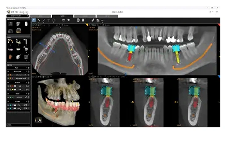
Foto: adoria.lv
Digital technologies have fundamentally changed the way dental implants are placed, making it more gentle for the patient and considerably more precise. Using 3D X-ray data, specialists can manufacture individual surgical guide templates that serve as navigation during the operation.
This means that the implant is placed at an angle and depth calculated virtually in advance, which maximises its stability and reduces the trauma to the surrounding tissue. Modern digital planning provides several essential advantages:
- A minimally invasive approach: thanks to a precise plan, the surgeon often does not need to make extensive incisions in the gum, which reduces post-operative swelling.
- A predictable result: the doctor sees, even before the procedure, how the implant will fit into the jaw, avoiding unforeseen anatomical shifts.
- Precise bone analysis: if the diagnostics show insufficient bone volume, bone augmentation is prescribed in a timely manner, in order to create a stable foundation for the new tooth root replacement.
- Integrated prosthetics planning: the 3D model makes it possible to coordinate the position of the implant with the placement of the future crown, ensuring flawless aesthetics and function.
When is additional preparation needed to strengthen the bone tissue?
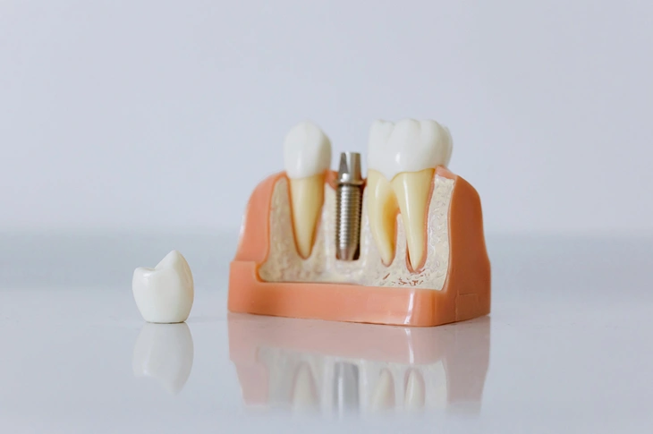
Foto: pexels.com/Pexels
In cases where the patient has lived without teeth for a long time, the jawbone naturally begins to shrink, or atrophy. If the 3D X-ray shows that the bone is too thin or low, preparation for dental implantation becomes a multi-stage process. The specialist may decide that bone grafting, or augmentation, or a sinus lift operation in the upper jaw, is needed first.
This stage is critically important, because dental implants cannot be placed "into a void" – they need stable support from all sides. Modern bone replacement methods make it possible to:
- Increase the thickness of the bone: using the patient's own tissue or synthetic replacement materials.
- Ensure durability: create conditions in which the implant will be able to withstand the full bite load for decades.
- An aesthetic result: a sufficient volume of bone and soft tissue ensures that the later dental prosthetics will look natural.
How does 3D diagnostics help to plan the next prosthetics stage?
A dental implant in itself is only an artificial root, but the patient's goal is the visible part of the tooth, which restores function and aesthetics. That is why dental prosthetics is planned simultaneously with the implantation, using the same 3D data. The doctor already sees from the outset what the shape of the crown will be and how it will fit into the existing row of teeth.
If, during the planning stage, the position of the future crown or prosthesis is not foreseen in time, the implant may be placed at an angle that later makes it difficult to fix the prosthesis aesthetically and stably. Only by combining detailed information about the anatomy of the jaw with the desired design of the row of teeth can the specialist guarantee that the new tooth will not function merely as a mechanical replacement, but will fit fully into the oral cavity, providing a natural visual appearance and a sense of bite.
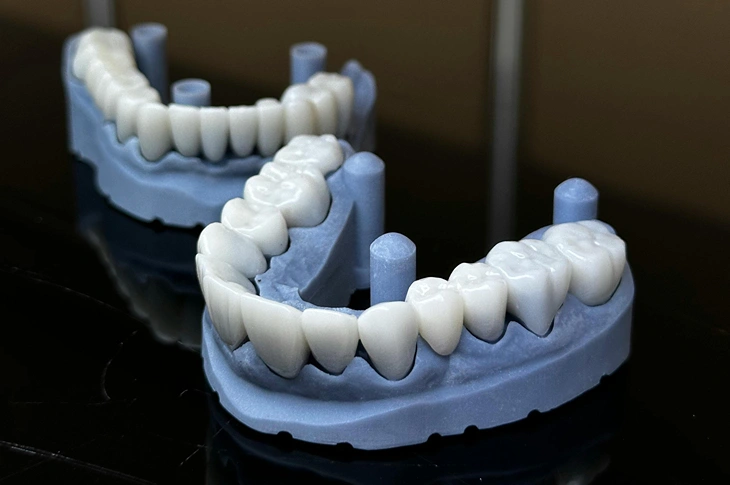
Foto: pexels.com/Pexels
What risks exist if implantation is performed without 3D diagnostics?
In modern dentistry, 3D X-ray diagnostics has become a mandatory standard, and in professional clinics the placement of dental implants is no longer planned without this examination. Attempts to save time or money at the expense of diagnostics can lead to serious medical complications, because without a three-dimensional overview the doctor operates "by feel", relying only on a superficial examination and an incomplete 2D image, in which the width of the bone is not visible.
When analysing cases in which negative reviews of dental implants are heard, the cause has most often been imprecise planning or unnoticed insufficiency of bone volume. The main risks of working without in-depth 3D data include:
- Chronic nerve irritation: if the implant is placed too close to the jaw nerve, a permanent tingling sensation or numbness in the lip, chin area and tongue may occur.
- Trauma to the sinuses: due to an incorrect implant angle, there is a risk of perforating, or creating a hole in, the wall of the paranasal sinus, which can cause chronic runny nose and infections.
- Implant rejection: if the volume of bone tissue has not been sufficient, the implant is unable to fully "grow in", or carry out osseointegration – the process in which living bone fuses tightly with the titanium screw.
- Trauma to adjacent teeth: in a 2D image it is impossible to see precisely the inclination of the roots of the adjacent teeth, so there is a risk of accidentally affecting them, causing damage to the adjacent teeth.
How long is the preparation stage until the operation itself?
To many patients it seems that diagnostics takes a long time, but modern digital processes are very efficient. Taking the 3D X-ray takes only a few minutes, and the image is immediately available on the specialist's computer for further analysis. Preparation for dental implantation itself, from the first consultation to the operation, can take from a few days to as much as several months, if prior treatment is needed.
If the patient's state of health is good and the bone volume is sufficient, the operation can follow soon after the diagnostics. However, if it is found that bone grafting is needed, patience becomes an important factor, because the formation of new bone takes time. It is essential to remember that haste is not an ally in implantology – careful examination and patient preparation of the tissue are the factors that ensure that the implant will serve as long as possible.
Frequently asked questions about 3D radiography and dental implantation
To dispel concerns, we will provide answers to questions that help to better understand the importance of 3D diagnostics.
- Is a 3D X-ray harmful to health?
Modern cone beam computed tomography (CBCT) devices use a very low radiation dose, comparable to the natural background radiation we receive every day. The benefit of precise operation planning incomparably exceeds the minimal radiation risk.
- How long is a 3D X-ray image valid?
Usually the image is current for approximately 6 months. If during this time no drastic changes have occurred in the oral cavity, for example no other teeth have been pulled, the specialist can use the existing data in order to carry out a precise placement of dental implants.
- Is a 3D X-ray also needed for replacing a single tooth?
Yes, even in the case of a single implant it is important to assess the width of the bone and the distance to the roots of the adjacent teeth. The individual anatomy of each patient is unique and unpredictable, and therefore the digital model is the only way to fully rule out the possibility of error and guarantee a safe course of the procedure.
Entrust the restoration of your smile to the Adoria specialists!
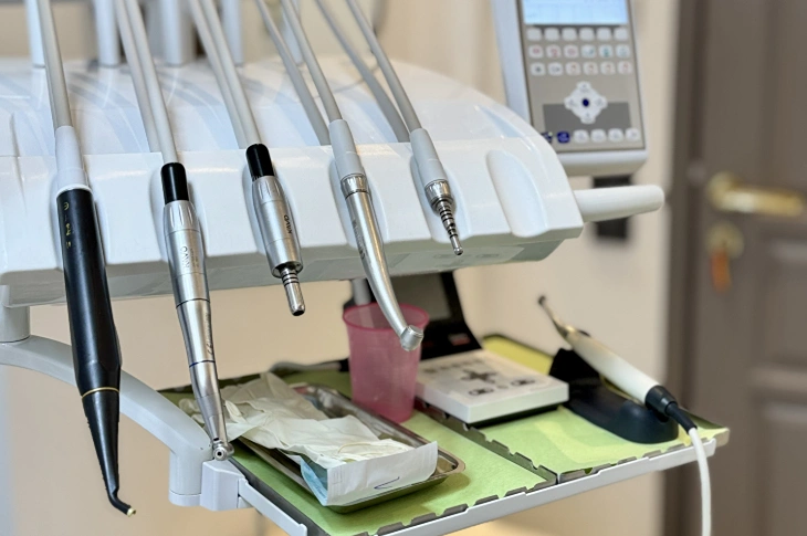
Foto: adoria.lv
To regain full chewing function and confidence in your smile, it is important to choose an experienced team and modern technologies. The Adoria Health and Beauty Centre offers full-cycle implantology, in which preparation for dental implantation takes place using the most precise 3D diagnostics and an individual approach to each patient. Book a consultation with our specialists by calling +371 67 315 000 or by filling in an application on the website, so that together we can create a safe path to your new smile.