3D X-ray vs 2D panoramic: which examination should you choose?
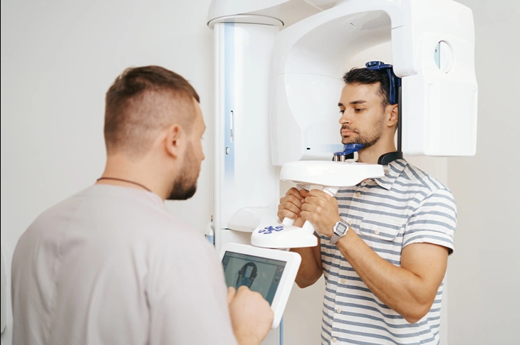
Foto: freepik.com/Freepik
In modern dentistry, precise diagnostics are the foundation of successful treatment, and the choice between different diagnostic methods can significantly affect the final result. Digital technologies offer both the traditional 2D image and a spatial analysis of the jaw structure, which helps the specialist to see the tiniest anatomical details. Darja Gvergžde, a dentist at the Adoria Health and Beauty Centre, explains in the following article in which cases a 3D dental X-ray is the best choice and how it differs from the usual panoramic image
In this article you will learn:
- The differences in technology: what the most essential differences are between a two-dimensional and a three-dimensional X-ray image.
- Diagnostic precision: in which cases a 3D dental X-ray is mandatory for precise and safe treatment.
- The course of the examination: how safe and fast is modern digital dental diagnostics?
What are the main differences between a 2D and a 3D image?
Traditional digital dental diagnostics is based on two-dimensional (2D) images, where the best-known method is the panoramic X-ray. Such an examination provides a clear, flat-format image in which both jaws, all the teeth and the sinuses are visible. Although this method is useful for assessing the general condition, it has a limitation – the structures in the image overlap, which makes it impossible to precisely determine, for example, the true depth of damage or the volume of bone.
By contrast, 3D technology, or the CBCT examination, makes it possible to obtain a spatial model that the doctor can view from any angle and in any cross-section. A 3D dental X-ray ensures millimetre precision, eliminating image distortions and the overlapping of structures and providing a complete picture of the oral cavity anatomy. To better understand the possibilities of both methods, it is worth comparing what the specialist sees in each of the images:
- 2D panoramic image: provides a general overview of the number of teeth, their arrangement, existing fillings, crowns and large caries lesions, and also makes it possible to assess the position of wisdom teeth and the condition of the sinuses.
- 3D digital model: reveals the precise density of the jawbone and exactly where the nerve canals, nerve plexuses and blood vessels are located, makes it possible to diagnose hidden root canals, microcracks in the tooth structure, and also to precisely determine the spread of an inflammatory process and the condition of the surrounding tissues. Such a detailed examination is especially significant when planning complicated procedures. For example, root canal treatment with a microscope is much more predictable if, even before starting the work, the doctor sees the entire canal system, which is not always possible to discern precisely on an ordinary X-ray.
In which cases is a panoramic X-ray sufficient?
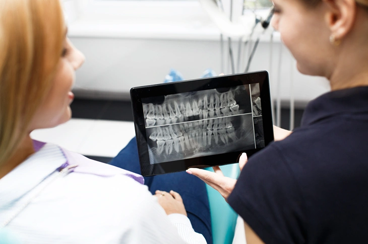
Foto: freepik.com/Freepik
Despite the rapid development of 3D technologies, the classic panoramic X-ray is still the primary diagnostic tool in dentistry. It is irreplaceable in annual preventive check-ups for detecting hidden caries lesions, changes caused by gum disease or the position of wisdom teeth. This method is fast, economically accessible and provides sufficient information for simpler dental procedures.
sufficient information for simpler dental procedures.
Specialists often use this examination to also notice pathological processes that are not visible to the naked eye, such as cysts or inflammations at the tooth roots. For more information about which diagnoses the doctor can make using a panoramic X-ray, we invite you to read the article "Hidden dental problems: what does the doctor see on a panoramic X-ray?".
The most essential reasons for choosing a 2D examination:
- General check-up: helps to assess the condition of all the teeth and the jaw joints at the same time.
- Orthodontics: necessary for children and adolescents in order to follow the development of the bite.
- Prevention: makes it possible to detect hidden caries lesions in good time, for example in the gaps between teeth.
- Operative planning of further treatment: serves as the basis for drawing up a further treatment plan.
When is a 3D X-ray examination mandatory?
In situations where maximum surgical precision is required, it is impossible to manage without spatial diagnostics. One of the most striking examples is the replacement of missing teeth, when dental implants are placed in the jawbone as the basis for further prosthetic work. During such a procedure, the dental surgeon must clearly see not only the thickness and height of the bone, but also the precise locations of the blood vessels and nerve plexuses, so that the manipulation is completely safe and predictable.
A 3D X-ray allows the specialist to virtually model each stage of treatment even before starting implantation and prosthetic work. To understand how digital technologies help in the course of dental implantology, the article "Preparing for dental implantation: why is a 3D study necessary?" provides useful information.
In addition to implantation, this method is also used in other cases:
- Orthodontics: helps to precisely determine the position of the tooth roots and the relationship of the jawbone, which is decisive when planning bite correction with braces or aligners.
- Complicated extractions: when planning the removal of wisdom teeth, the spatial data help to notice the curves of the roots and their proximity to the main facial nerves, significantly reducing the risk of trauma.
- Endodontic retreatment: in cases where repeated cleaning of the canals is necessary, the image reveals previously unnoticed anatomical details or hidden foci of inflammation.
- Restoration of bone volume: makes it possible to precisely calculate the amount and shape of missing bone tissue before its augmentation surgery.
Foto: freepik.com/Freepik
What you need to know about safety and radiation doses?
For many patients, the question of how great the impact of such an examination is on the body causes concern. It must be emphasised that modern digital dental diagnostics ensures the minimum possible radiation dose. Using pulsed radiation technology, the device reads the data in just a few seconds, as a result of which the radiation dose received is comparable to the natural background that a person receives daily from the surrounding environment or during a flight by aeroplane.
Therefore, a 3D dental X-ray is permissible for both children and pregnant women, if there are justified indications. Digital systems automatically adjust the intensity of the beam to the patient's body build, ensuring the highest quality image with minimal exposure. A precise examination protects against errors in diagnosis, which ultimately spares the patient's health and prevents unnecessary manipulations or the risk of complications.
Aspects of diagnostic safety:
- Minimised time: the exposure lasts fractions of a second, reducing the load on the body.
- Precise focus: the radiation is directed only at the necessary examination field.
- Digital processing: the obtained data are stored electronically, reducing the need for repeated images.
How much do diagnostic services cost?
When planning a treatment budget, an essential factor for patients is the current dental X-ray prices. Usually a 2D panoramic image is more financially accessible, because it requires less time for data analysis and is carried out with simpler equipment. Spatial diagnostics, on the other hand, requires greater technological resources and the time of a highly qualified specialist for interpreting the image, which is why its cost is higher.
However, it is important to weigh that a 3D, or CBCT, examination provides incomparably broader information. An investment in precise diagnostics at the initial stage can protect against unforeseen costs later, because it makes it possible to avoid unsuccessful treatment or complications that could arise due to insufficient information.
A modern dental diagnostics office in the centre of Riga
Digital diagnostics and three-dimensional examinations are today the gold standard, which helps to ensure a safe and predictable treatment result. The latest-generation technologies available at the Adoria Health and Beauty Centre make it possible to take both 2D panoramic X-ray images and an in-depth 3D study in a calm and patient-friendly way.
If high-level digital dental diagnostics in the centre of Riga are needed, the Adoria Health and Beauty Centre offers immediate help and an individual approach to each patient. Our specialists will promptly assess the situation, using the most modern equipment, in order to draw up the treatment plan most suitable for you. Book an appointment with our experienced professionals by calling +371 67 315 000 or by filling in the application on the website, and receive quality care in a modern environment!