
Hidden dental problems: What does a dentist see on a panoramic X-ray?
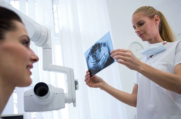
Photo: magnific.com/Magnific
Do you know the feeling when your teeth seem healthy, nothing hurts, and you follow your daily hygiene routine perfectly, yet a visit to the dentist reveals problems that require immediate intervention? Often, even an experienced specialist cannot see during a visual examination what is happening beneath the gums or in the thickest layer of tooth enamel. In such cases, precise imaging examinations play a decisive role, helping to detect structural changes in the jawbone before they cause acute pain or tooth loss.
As the certified dentists at the Adoria health and aesthetics clinic point out, precise dental diagnostics in Riga is the foundation of successful and gentle treatment. A panoramic X-ray is one of the most important tools that allows the specialist to assess the overall health of the oral cavity, revealing both unnoticed tooth decay and serious tissue damage such as dental cysts, thus ensuring timely treatment.
What is a panoramic X-ray?
A panoramic X-ray, or orthopantomogram, is a two-dimensional (2D) imaging method that provides a complete overview of the entire oral cavity, including the upper jaw, lower jaw, all teeth, their roots and the temporomandibular (jaw) joints. Unlike a local X-ray, which shows only one or two adjacent teeth, this examination generates a broad map of the anatomical structure. This basic tool of general dentistry allows the doctor to assess bone density and detect pathologies hidden deep in the tissues that are not visible to the naked eye.
The most common hidden pathologies found in imaging
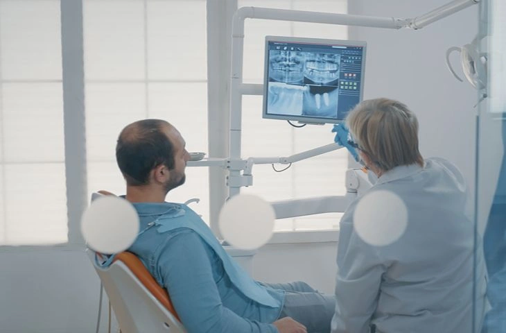
Photo: magnific.com/Magnific
Clinical practice has shown that even a visually intact tooth can hide deep and progressive damage. Imaging most often reveals problems such as:
Hidden decay in the spaces between teeth
One of the most common problems, and one that is almost impossible to notice during a standard examination, is hidden tooth decay. It primarily forms between two adjacent teeth (on the contact surfaces) or under old fillings and dental crowns. Since the outer layer of the tooth (the enamel) may look healthy on the surface, the damage develops for a long time in the dentin – the inner, softer layer of the tooth. On the X-ray, these damaged areas appear as darker spots or shadows. When these changes are detected in time, early dental therapy is prescribed, stopping further breakdown of the tooth structure and preventing inflammation of the nerve.
Root canal inflammation
If tooth decay is left untreated for a long time, bacteria penetrate the dental pulp (the living tissue of nerves and blood vessels), causing an infection. The X-ray image often shows that the patient has developed root canal inflammation. It is diagnosed as a dark, altered area in the jawbone around the tip of the tooth root. Even if the person does not feel significant pain in daily life, this process indicates chronic inflammation. In such situations, endodontic intervention is needed – mechanical cleaning and hermetic filling of the root canals.
Dental cyst – an unnoticed threat to the jawbone
Another serious and clinically significant problem is a dental cyst – a pathological sac filled with tissue fluid, which usually forms as a result of infection at the root tip of a dead tooth. It is the body's natural defensive reaction, an attempt to isolate the source of infection from healthy bone tissue. Cysts grow gradually and may cause no physical symptoms for years, but over time they destroy the surrounding jawbone and can put pressure on the roots of adjacent teeth. On an X-ray, they appear as well-defined round or oval shadows. Treatment of this pathology most often involves dental surgery to safely remove the lesion while trying to preserve the functionality of the natural tooth.
Periodontitis and bone loss
Periodontitis is a complex inflammation of the gums and the supporting structures of the teeth, resulting in irreversible breakdown of the jawbone. On a panoramic image, the doctor can see exactly how far the bone level has receded around the tooth roots. This information helps determine the stage of the disease and develop a targeted treatment plan to stop bone loss and prevent teeth from becoming loose.
Overview of symptoms and hidden causes
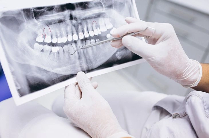
Photo: magnific.com/Magnific
To make the processes in the oral cavity easier to understand, the most common indications and their explanations from a diagnostic point of view are summarised below:
| Patient complaints (Symptoms) | Possible cause - on the X-ray | Solution at the clinic |
|---|---|---|
| Temporary sensitivity to cold/sweet, no visible cavity | Hidden decay between teeth | Dental therapy and fillings |
| Pain when biting, swollen gums near the root | Root canal inflammation | Endodontics (root canal treatment) |
| No complaints, but facial asymmetry or swelling | Dental cyst or granuloma | Dental surgery, removal of the lesion |
| Bleeding gums, teeth that look "longer" | Periodontitis, bone loss | Deep cleaning, periodontist care |
Frequently asked questions about dental imaging
How often should a panoramic X-ray be taken?
For preventive purposes, this examination is recommended once every one to two years. The interval depends directly on the condition of the patient's oral cavity, previous diseases and planned procedures (for example, before orthodontic treatment or implant placement).
Is this examination safe for health?
Modern digital X-ray equipment uses a minimal radiation dose, many times lower than older analogue devices. The examination is completely safe; in case of an existing or possible pregnancy, the attending doctor or X-ray technician must always be informed in advance.
How does a panoramic image differ from a 3D scan?
To learn more about the detailed differences between 2D and 3D technologies, we invite you to read our dedicated article: 3D X-ray vs 2D panoramic: Which examination should you choose?.
Take a step towards a healthy smile
The health of your oral cavity is closely linked to the overall well-being of your body. Even if you do not feel any discomfort right now, a preventive check-up and timely imaging examinations can save you both time and considerable money in the future by preventing serious diseases from developing. Take control of your health today – diagnostics is the first step towards a long-term result.
Book an in-person appointment with our highly qualified specialists at Adoria by calling +371 67 315 000 or by conveniently filling in the online booking form on our website!